Non Destructive Testing and Imaging of Cosmetics and Personal Care Products

Our non destructive testing systems using x-ray ct scanning or IR can screen for defects in multi component products without the need for opening, allowing you a unique view into components and how they integrate together, or where stress points can arise. We can help you discover breakage points for usage or shipping, or highlight closure and seals for liquid containers or airtight packaging needs.
Featured Applications
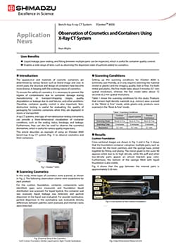
Observation of Cosmetics and Containers Using X-Ray CT System
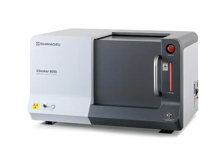
A non-destructive approach to visualizing cosmetic products and their packaging with advanced X-ray imaging technology. This study explores a wide range of cosmetic formulations and material components, including creams, emulsions, and container structures, enabling detailed observation of internal features such as air bubbles, phase distribution, and structural defects without altering the sample. An X-ray computed tomography (CT) was use to generate high-resolution, three-dimensional images of both cosmetic contents and their containers. Analysis is performed using the Shimadzu XSeeker 8000 Benchtop Microfocus X-Ray CT System, which enables precise internal imaging under controlled scanning conditions without sample preparation or destruction. This allows simultaneous evaluation of formulation homogeneity and container compatibility as the platform provides detailed 3D visualization of internal structures in a fully non-destructive manner. The process enables rapid defect detection, formulation optimization, and comprehensive product evaluation in a single workflow.
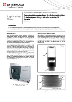
Example of Observing Resin Bottle Containing Hair Coloring Agent Using a Microfocus X-Ray CT System
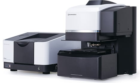
Non-destructive imaging provides a perspective on cosmetic packaging and formulation quality to support leak prevention, formulation integrity, and container compatibility, directly impacting consumer product safety, shelf stability, and customer satisfaction. This study examines hair coloring agents within resin bottles, focusing on the physical distribution of the formulation, as well as structural features such as air bubbles, gaps between bottle and cap, and internal filling uniformity. Microfocus X-ray computed tomography (CT) to capture detailed internal structures in three dimensions. Analysis is performed using the Shimadzu inspeXio™ SMX™ 225CT FPD HR Plus Microfocus X-ray CT system, enabling high-resolution fluoroscopic, cross-sectional, and 3D imaging of both the container and its contents. The technique allows precise visualization of interfaces, internal defects, and liquid distribution without physically altering the sample. Complete, non-destructive visualization of internal structures enables rapid detection of defects such as leakage paths, voids, and assembly inconsistencies to drive improved product quality, packaging reliability, and formulation performance.
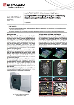
Example of Observing Paper Diaper and Sanitary Napkin Using a Microfocus X-Ray CT System
Non destructive imaging is used to investigate the internal structure of absorbent systems composed of superabsorbent polymers (SAP) and cotton like pulp matrices, which are key functional components responsible for liquid uptake and retention. The methodology employs microfocus X-ray computed tomography (CT) to visualize internal structures in three dimensions before and after liquid absorption. Analysis is conducted using the Shimadzu inspeXio™ SMX™ 225CT FPD HR Plus Microfocus X-ray CT system, generating high-resolution fluoroscopic, cross-sectional, and 3D images. This enables direct observation of SAP particle distribution, expansion upon absorption, and water retention pathways within the material matrix under controlled imaging conditions. Real-time, non-destructive visualization of structural changes within the same sample, provides access to precise evaluation of absorption mechanisms and supports the development of more effective, high-performance personal care products.
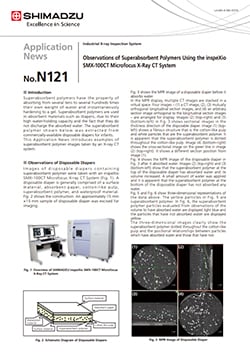
Observations of Superabsorbent Polymers Using the inspeXio SMX-100CT Microfocus X-Ray CT System
Superabsorbent polymers (SAPs) embedded within cotton-like pulp matrices are key components responsible for high-capacity liquid absorption and retention in consumer formulations. Understanding the distribution, morphology, and swelling behavior of these polymers is essential for personal care and cosmetic product development, where absorbent systems influence texture, performance, and user experience in products such as pads, wipes, and hybrid formulations. The methodology utilizes microfocus X-ray computed tomography (CT) to non-destructively visualize internal structures in both dry and liquid-absorbed states. Analysis is performed using the Shimadzu inspeXio™ SMX 100CT Microfocus X-ray CT system, generating high-resolution cross-sectional and three-dimensional images without physical sectioning or complex sample preparation. Controlled imaging conditions enable detailed observation of particle size (tens to hundreds of micrometers), spatial distribution, and volumetric expansion after absorption. True non-destructive, high-resolution 3D visualization, allows direct comparison of structural changes before and after absorption empowering deeper understanding and optimization of advanced absorbent systems.
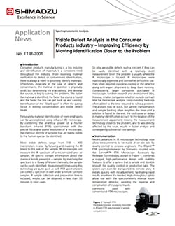
Visible Defect Analysis in the Consumer Products Industry – Improving Efficiency by Moving Identification Closer to the Problem
Consumer products manufacturing is a big industry and identification of materials is a consistent need throughout the industry. From incoming material verification to defect or contaminant identification, there is always a need to positively identify materials. Oftentimes, especially in the case of defects and contaminants, the material in question is physically small, but determining the true identity, and likewise the source, is key to solving the problem. The faster the material is identified, the faster the source is found and the faster the line can get back up and running. Identification of the “black spot” is often the gating factor in solving contamination and visible defect issues. Fortunately, material identification of even small spots can be accomplished using infrared (IR) microscopy. By combining the analytical power of a Fourier transform infrared (FTIR) spectrometer with the precise focus and spatial resolution of a microscope, the chemical identity of samples that are barely visible to the human eye can be identified.