AAPS Pharm Sci 360 2026 (American Association of Pharmaceutical Scientists)
October 26-28
Ernest Morial Convention Center
New Orleans, LA
Booth #1204
The bases and nucleotides are generally separated by ion-exchange or reverse-phase mode HPLC and detected by UV absorbance detection.
In the example introduced here, nucleic acid-related compounds were analyzed by LC/MS, which offers mass information and enables high-sensitivity analysis.
Fig. 1 shows the structure and ESI mass spectrum of the purine base adenine and of the nucleotide adenosine. Under acidic conditions in the positive ion mode, the protonated molecule [M+H]+ is observed as a reference peak.
Fig. 2 shows the LC/MS analysis results of a standard mixture of nucleic acids. SIM measurements were conducted using [M+H]+ as the detected ion for each nucleic acid. Satisfactory separation of the 15 components was achieved.
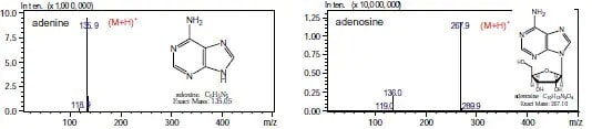
Fig. 1 Positive-Ion ESI Mass Spectrum of Adenine and Adenosine
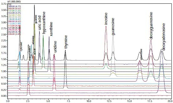
Fig. 2 SIM Chromatogram of Standard Mixture of Nucleic Acid Bases and Nucleotides
AAPS Pharm Sci 360 2026 (American Association of Pharmaceutical Scientists)
October 26-28
Ernest Morial Convention Center
New Orleans, LA
Booth #1204
ASMS (American Society for Mass Spectrometry) 2026
May 31 - June 4
San Diego Convention Center
San Diego, CA
Suite 5, 6
Pharma Community Network Event
Shimadzu Scientific Instruments and ZefSci invite you and your analytical teams to a pharma community networking event to celebrate 150 years of science and innovation at The Foundry & Lux in South San Francisco. Our event will be built around innovation, collaboration, and connection.
Shimadzu Scientific Instruments Opens Boston Location of Its R&D Center Focus will be on promoting customer-oriented development to expand business in the pharmaceutical field
Shimadzu Scientific Instruments, Inc. (SSI, Columbia, Maryland, USA), a Shimadzu Group company, has opened a satellite lab in Boston, Massachusetts to be the base of its collaborative research and development activities on the East Coast. Established to conduct research and development more closely linked to customers, SSI's R&D Center consists of three bases, with the main facilities at its Maryland headquarters, a West Coast location, and this new space in Boston near the city center. The Boston lab was set up by partnering with Labshares, a shared laboratory service provider for life science companies.