Thin Film Sample Measurement Methods and Precautions
Various measurement methods are available in infrared spectrophotometry, and appropriate use of these depends on the measurement object (sample), analytical objective and other factors. For measurement of thin film samples with a film thickness of up to 1 µm, the high-sensitivity reflection method is widely used, but recently, single-reflection ATR is also being utilized. This article introduces examples of thin film analysis using the high-sensitivity reflection and single-reflection ATR methods.
1. Measurement of Thin Film on Metal
Reflection absorption spectroscopy (RAS) is suitable for measurement of thin film on a metallic substrate. RAS is an external reflection (specular reflection method) method in which the angle of incidence is 70° or greater, whereby the substrate-associated peak vibration in the perpendicular direction is intensified. Using this, it becomes possible to measure thin film spectra that cannot be measured by the specular reflection method using an approximately 10° incidence angle.
Fig. 1 shows a reflection absorption spectrum of a mono-layer fibrinogen film on metal plating. Measurement was conducted at an 80° incidence angle. Fibrinogen, a water soluble protein present in blood, is a rod-like molecule with a molecular weight of about 340,000, a diameter of 9 nm, and length of about 45 nm. Amide I and amide II in the vicinity of 1666 and 1545 cm-1, respectively, and the stretching vibration of N-H in the vicinity of 3312 cm-1 are clearly confirmed.
Fig. 2 shows a reflection absorption spectrum of a multi-layer sample (gold / hydroxyapatite / fibrinogen) consisting of a gold plating substrate, a hydroxyapatite layer (thickness about 10 nm) and a mono-layer fibrinogen film. The hydroxyapatite peak and fibrinogen peak are clearly seen in the vicinity of 1100 cm-1. In the measurement of this sample, the infrared light irradiated on the sample was reflected from the fibrinogen surface as well as the interface between the fibrinogen and hydroxyapatite layers. However, compared to the reflected light from the gold, that amount is extremely small, leading to the assumption that the obtained spectrum is derived from light reflected off the gold surface after having been transmitted through the fibrinogen and hydroxyapatite layers. The analytical conditions used for the spectra in Figs. 1 and 2 are shown in Table 1.
Table 1: Analytical Conditions 1
| Accessory : VeeMAX Incidence angle : 80 Resolution : 4 cm-1 Number of scans : 400 Detector : MCT |
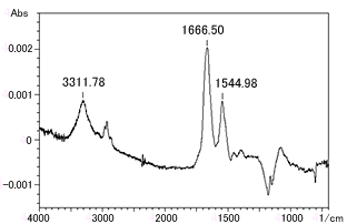
Fig. 1 Reflection Absorption Spectrum of Fibrinogen Thin Film on Gold Plating
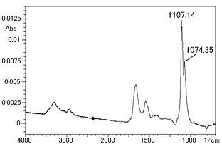
Fig. 2 Reflection Absorption Spectrum of Hydroxyapatite / Fibrinogen Multi-Layer Thin Film on Gold Plating
2. Precautions in Reflection Absorption Spectroscopy (RAS)
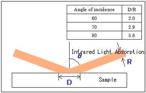
Fig. 3 Spreading of Light Beam in High-sensitivity Reflection Method (D/R is calculated assuming parallel light beams)
- Because a large angle of incidence is used in Reflection absorption spectroscopy (RAS), the light beam spreads widely on the metal surface (see Fig. 3).
When the measurement sample is smaller than the light beam, a mask is used to eliminate reflected light other than that from the sample. The greater the angle of incidence, the higher the sensitivity that can be expected; however, since this is accompanied by wider spreading of the light beam, the light directed at the detector is reduced. Reducing the amount of light detected will result in increased noise. Therefore, when using RAS, it is essential to select the angle of incidence according to the sample and analytical objective. Now, when conducting analysis with a large angle of incidence and a small amount of light, an MCT detector is effective. - The mask used when the measurement sample is smaller than the light beam has a black coating on its surface to impart low reflectivity. However, as this reflectivity is not zero, there may be some influence due to reflected light from the surface of the mask when using Reflection absorption spectroscopy (RAS). This influence can be eliminated by applying soot to the surface of the mask to reduce the reflectance to zero. Application of soot is often performed using a candle.
- RAS is often conducted in combination with a grid polarizer. Since only parallel polarized light contributes to the above-mentioned "intensification of the metal substrate-associated peak that is vibrating in the perpendicular direction", the polarizer is used to cut the vertical polarized light, which has no effect on the peak, thereby giving the appearance of increased peak intensity. However, since use of the grid polarizer reduces the amount of light to less than one half, there is an increase in noise. In RAS, it is not absolutely necessary to use a grid polarizer.
(For details on reflection absorption spectroscopy (RAS), refer to "Fundamentals of FTIR - ABCs of Reflection Absorption Spectroscopy".)
3. Measurement of Thin Film on Non-Metallic Substrate
Reflection absorption spectroscopy (RAS) is extremely effective for measurement of thin films on metallic substrates; however, this method is unsuitable for measurement of thin films on non-metallic substrates such as glass, silicon wafer and plastics.
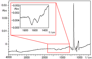
Fig. 4 Reflection Absorption Spectroscopy (RAS) of Fibrinogen Thin Film on Hydroxyapatite
Fig. 4 shows the reflection absorption spectrum of a fibrinogen mono-layer film on a mirror-polished hydroxyapatite sintered plate. The inset in Fig. 4 is a magnification of the range from 1900 to 1300 cm-1. As the reflectivity of hydroxyapatite is lower than that of metal, the "intensification of the peak that is vibrating in the perpendicular direction due to parallel polarized light" is not seen. Furthermore, the amide I and amide II peaks are inverted due to the influence of reflection from the fibrinogen film surface. Although peak shapes are apparent in the vicinity of 1250 to 1000 cm-1, these are the residues of the hydroxyapatite reflectance spectrum.
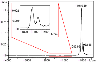
Fig. 5 ATR Spectrum of Fibrinogen Thin Film on Hydroxyapatite
Fig. 5 shows an ATR spectrum of the same sample measured by single-reflection ATR. A Ge prism was used. The peaks in the vicinity of 1084, 1016 and 962 cm-1 are peaks derived from hydroxyapatite. The inset in Fig. 5 is a magnification of the range from 1900 to 1300 cm-1. The inverted amide I and amide II peaks seen in Reflection absorption spectroscopy (RAS) are obtained normally in this spectrum.
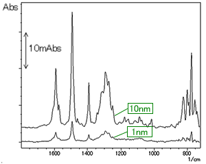
Fig. 6 ATR Spectra of NPD Thin Film on ITO Film (Upper: 10 nm, Lower: 1 nm)
Fig. 6 shows the ATR spectra of N, N'-di (1-naphthyl)-N, N'-diphenylbenzene (NPD) thin film measured by the single-reflection ATR method (Ge prism) (after subtraction with ITO spectrum). NPD is a substance known as a material of the electron hole transport layer in organic EL displays, and the NPD film thicknesses (measured here) are 1 nm and 10 nm. A good spectrum was obtained even with the 1 nm thickness.
The analytical conditions used for the spectra in Figs. 5 and 6 are shown in Table 2.
Table 2: Analytical Conditions 2
| Accessory : MIRacle Prism : Ge Resolution : 4 cm-1 Number of scans : 100 Detector : DLATGS |
Fig. 6 ATR Spectra of NPD Thin Film on ITO Film
(Upper: 10 nm, Lower: 1 nm)
- In ATR methods, measurement is contacted with the sample tightly pressed against the prism, and the spectrum is influenced by the contact condition. In particular, if the state of contact is poor when measuring thin film, a spectrum may not be obtained. Therefore, it is necessary to perform measurement after ensuring that the contact surfaces of the sample and prism are clean and without any adhering substances. Furthermore, if the substrate has a rough surface, contact will be poor, and good results will probably not be obtained.
- Theoretically, better results with higher sensitivity can be obtained with multi-reflection ATR as compared to single-reflection ATR due to the strong intensity of peaks generated through a greater number of reflections. In actuality, however, it is common to obtain peaks by single-reflection ATR that cannot be obtained using multi-refection ATR. This is because the prism used in multi-reflection ATR is large, and minute particles of dirt and other adhering substances from the sample can easily create a gap to make it difficult to obtain good contact with the prism. In single-reflection ATR, the prism is small, and even if there are minute particles on the surface of the sample, they can be avoided to allow good contact.
5. Conclusion
This FTIR Talk Letter introduced Reflection absorption spectroscopy (RAS) and the single-reflection ATR technique as methods for measuring thin film samples. RAS is unsuitable when the substrate is non-metallic, but thin film on a metallic substrate can be easily measured in a non-contact state. On the other hand, single-reflection ATR requires that the sample and prism be in close contact; however, it is extremely effective for measurement of thin films on non-metallic substrates.
Of the measurement examples introduced in this article, the samples for Figs. 1 to 5 were provided by Mr. Ikoma and Mr. Monkawa of the Biomaterials Center, National Institute for Materials Science, Japan. The sample for Fig. 6 was provided by Mr. Ide of the Optoelectronic Industry and Technology Development Association Yamagata Research Center, and Shiroto Research Laboratory, Faculty of Engineering, Yamagata University, Japan.