AABC (Advanced Automotive Battery Conference) 2025
December 9-11
Caesars Palace
Las Vegas, NV

Shimadzu offers a complete range of instrumentation to support advancements in the field of biomaterials and new medical treatments, including regenerative tissue implants, orthopedics, drug delivery, and medical devices. Mechanical test equipment such as universal test machines and fatigue testers enable the evaluation of the biomechanical and physical properties of biological samples, tissue, biomaterials, elastomers, polymers, metals, and shape memory alloys. They can also be used to study the suitability and reliability of medical devices and packaging designs. Our advanced x-ray imaging systems can be used for the inspection of animal specimens and tissue used in experimental research, and our particle size analysis and life science instrumentation offer innovative solutions for the analysis of biological
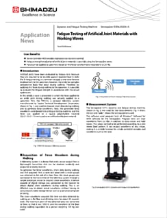
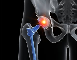
In this article, a case is presented in which the force applied to an ankle joint during walking was actually applied to a specimen. First, the TM5135, a compact telemetry system manufactured by Toyota Technical Development Corporation shown in Fig. 1, was used to measure strain data during walking and to generate force waveforms. Next, the generated force waveforms were loaded into a fatigue testing machine, and the force was applied to a square polyethylene material (23.5×23.5×12mm) used as an artificial ankle joint material.
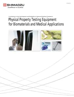
To guarantee the safety and efficacy of medical equipment and technologies in the wake of medical breakthroughs, more active efforts are being made to reinforce the evaluation criteria and the regulations governing the standardization of measuring instruments and test methods. Medical equipment manufacturers and research organizations around the world are conducting research and development into medical equipment based on mechanical properties evaluation and finite-element analysis. Shimadzu Corporation is applying the technical expertise cultivated through physical testing, quality control, and full-scale testing in materials development to the fields of leading-edge medicine and biomaterials evaluation.
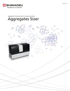
The Aggregates Sizer aggregation analysis system enables the quantitative evaluation of particle amounts in the 0.1 μm to 10 μm range as a concentration (units: μg/mL or particles/mL). Aggregations of biopharmaceuticals can be categorized into 4 ranges: nanometer (<100 nm), submicrometer (100 nm to 1 μm), subvisible (1 to 100 μm), and visible (>100 μm), according to their particle size. Until now, particles in the submicrometer to subvisible portion of this range (100 nm to 10 μm) were generally measured by combining multiple methods.
The inspeXio SMX-100CT system has a micro focus X-ray generator (max.100kV) and high sensitive image intensifier, making this system useful for the observation of soft materials (resin, bone etc.). The internal structures of small insects such as internal muscles and nerves can be observed nondestructively by CT imaging.
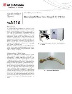
At universities, pharmaceutical manufacturing companies and in other research fields, a variety of research using experimental animals is being conducted, including research into bone diseases, as well as drug administration evaluations, and fat measurements for metabolic research. The main experimental animals are rats, mice, and rabbits. In this context, X-ray CT systems are often used to observe and analyze the bones of small animals, and for research on the teeth of humans and small animals. Observations of small animals consists of in vivo CT imaging of living animals, and in vitro CT imaging of dead animals and excised parts. This article introduces imaging (in vitro) CT data from mouse femurs, taken using the inspeXio SMX-100CT, as well as analytical results from 3-dimensional analysis software utilizing the CT data.