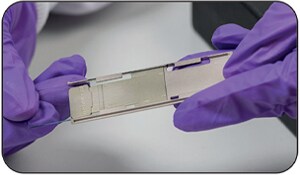
FlexiVision-mini ITO slide used with Adaption-miniadapter during analysis
The Shimadzu Benchtop Imaging Kit contains all the components necessary to conduct imaging experiments on your Shimadzu MALDI Benchtop Instrument (not including the MALDImini).
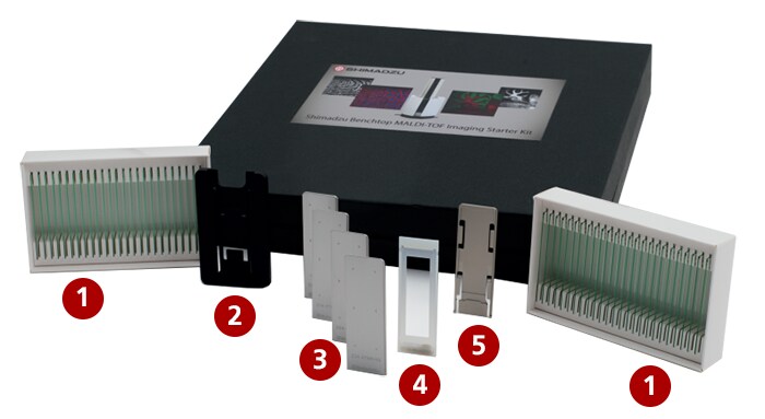
| 1 | FlexiVision™-mini ITO slides (2 packs of 25 slides) |
Single-use glass slides custom-sized to fit in MALDI-8020™/ MALDI-8030™ mass spectrometers when assembled with Adaption™-mini glass slide adapter |
| 2 | Adaption-mini carrier | Used during optical scanning to securely hold either the Adaption-mini adapter/FlexiVision-mini ITO sample slide or the FlexiMass™-SR1 Stainless steel sample plate. Ensures the sample slide surface does not contact the scanner surface |
| 3 | FlexiMass-SR1 reusable metal sample plates (4 plates) | Reusable* FlexiMass format, engraved with the accessible sample area to aid mounting of samples |
| 4 | Slide mask | Fits over the FlexiVision-mini ITO slides during matrix coating or tissue mounting to clearly identify the usable target area |
| 5 | Adaption-mini ITO glass slide adapter | Custom glass slide holder for use with the FlexiVision-mini ITO slides |
| MALDI Solutions™ imaging acquisition license | Provides the imaging acquisition wizard to co-register the optical image and define the area to be acquired | |
| IonView™ MALDI imaging software license | Software for processing MALDI images. Reads data directly from MALDI Solutions |
* follow appropriate washing procedure.
FlexiVision-mini ITO slide used with Adaption-miniadapter during analysis
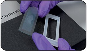
FlexiVision-mini ITO slide used with Slide Mask duringmatrix coating
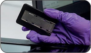
FlexiVision-mini ITO slide (within Adaption-miniadapter) used with Carrier during optical scanning
The MALDI Solutions IonView software (included in kit) provides an easy workflow for routine processing of MALDI images such as snapshots, overlays and displays of regions of interest and pixel spectra.
| Sample | Compounds in fingermark |
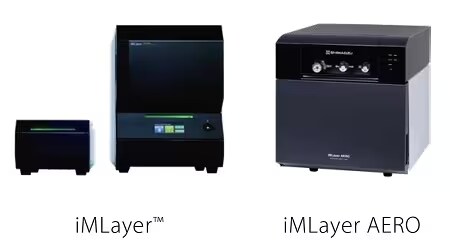
| Matrix | DHB, sublimated with Shimadzu iMLayer device |
| Measurement region | 25,600 pixels |
| Measurement time | Around 3.5 hours |
| Experiment details | Laser repetition rate of 200 Hz and 100 shots per pixel |
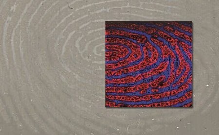
Overlay of m/z 381 and m/z 384
The workflow is simple making it amenable to new users learning the technique as well as experienced imaging users.
There are three main steps in the MALDI imaging workflow after collection of tissue sections:
Solutions are provided in each step to make an otherwise typically difficult imaging workflow into a simple, user-friendly application with a successful outcome for your laboratory.
* Optional Item
* Not included in Benchtop MALDI-TOF Imaging Starter kit
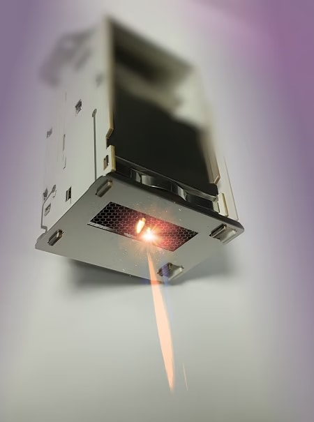
The AeonDetector has up to 7x longer life and reduced aging rate.
The Shimadzu imaging benchtop MALDI-TOF solution has design features well-suited to a demanding application such as MALDI imaging:
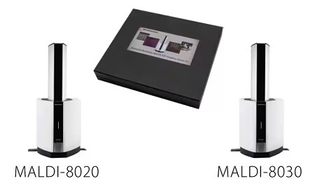
Class-leading sensitivity and mass resolution of the MALDI-8020 and MALDI-8030.
FastMS feature: the combination of the 200 Hz laser, fast sample introduction (< 3mins) and quick sample stage provides a quick analytical turnaround time (speed of up to 15 pixels per second, based on lipid imaging acquisition with mass range m/z 100-1200, 50 μm spacing and an accumulation rate of 10 shots/profile)
Robustness provided by the class-leading long lasting laser lifetime of 2 billion shots, the self-cleaning ion optics using the patented TrueClean one-click laser technology, and the longer-life AeonDetector
.